Understanding the Inner Workings of Spider Silk: A Leap Toward Synthetic Production
Unraveling the secrets of spider silk and its awe-inspiring properties has been a quest for many scientists. Its lightness, strength, and flexibility make it a coveted material, which, if artificially replicated, could redefine industries and replace Kevlar, polyester, and carbon fiber

Image by Canva
Unraveling the secrets of spider silk and its awe-inspiring properties has been a quest for many scientists. Its lightness, strength, and flexibility make it a coveted material, which, if artificially replicated, could redefine industries and replace Kevlar, polyester, and carbon fiber. Despite significant efforts, this arachnid craftsmanship remains a marvel of nature that has yet to be authentically duplicated.
One hopeful contender in this fascinating race is Irina Iachina, a postdoctoral biophysicist from the University of Southern Denmark (SDU). Iachina's fascination with spider silk has led her from her master's studies at SDU to her current research at the Massachusetts Institute of Technology in Boston, funded by the Villum Foundation.
In a bid to demystify the microscopic architecture of spider silk, Iachina has teamed up with Jonathan Brewer, an associate professor, and biophysicist at SDU. An expert in probing biological structures using microscopes, Brewer is pivotal to their collaborative endeavor. Their innovative work, a world's first, involves studying the interior parts of spider silk using an optical microscope without any physical alterations to the silk. Their study was published recently in both the Scientific Reports and Scanning journals.
In a statement, Brewer shared, "We have used several advanced microscopy techniques, and we have also developed a new kind of optical microscope that allows us to look all the way into a piece of fiber and see what's inside."
Prior methods of analyzing spider silk provided important insights but presented challenges, as they required opening the silk fiber for microscopic examination or freezing the samples, which could potentially modify the silk's structure. To circumvent these issues, Iachina expressed, "We wanted to study pure and unmanipulated fibers that have not been cut, frozen, or manipulated in any way."
To achieve this, the researchers employed non-invasive techniques like Coherent Anti-Stokes Raman Scattering, Confocal Microscopy, Ultra-resolution Confocal Reflection Fluorescence Depletion Microscopy, Scanning Helium Ion Microscopy, and Helium Ion Sputtering.
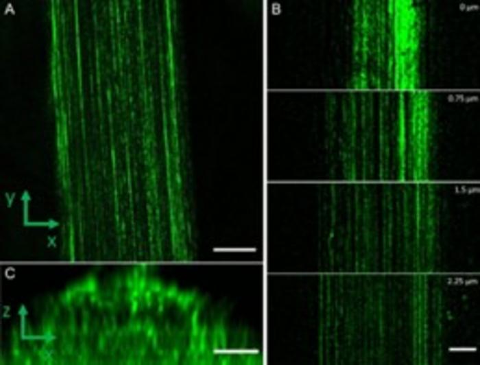
“Novel imaging techniques reveal new insights into the macro- and nanoscopic structure of Major (MAS) and Minor (MiS) Ampullate silk fibers from pristine samples of the orb-web spider Nephila madagascariensis,” the authors wrote. “Untreated threads were imaged using Coherent Anti-Stokes Raman Scattering and Confocal Microscopy, which revealed an outer lipid layer surrounding an autofluorescent protein core that is divided into two layers in both fiber types. Helium ion imaging shows the inner fibrils without chemical or mechanical modifications. The fibrils are arranged parallel to the long axis of the fibers with typical spacing between fibrils of 230 nm ± 22 nm in the MAS fibers and 99 nm ± 24 nm in the MiS fibers. Confocal Reflection Fluorescence Depletion (CRFD) microscopy imaged these nano-fibrils through the whole fiber and showed diameters of 145 nm ± 18 nm and 116 nm ± 12 nm for MAS and MiS, respectively.”
Their research revealed the complex architecture of a spider's silk fiber. It comprises two outer lipid layers with numerous straight, closely-packed fibrils. These fibrils range between 100 and 150 nanometers, which is smaller than what a regular light microscope can measure.
In a surprising revelation, Iachina noted, "They are not twisted, which one might have imagined, so now we know that there is no need to twist them when attempting to create synthetic spider silk."
Their exploration focused on the golden orb-web spider, Nephila madagascariensis, which generates two silk types: MAS (Major Ampullate Silk fibers), used to construct the spider's web and serve as a lifeline, and MiS (Minor Ampullate Silk fibers), a more elastic auxiliary construction material.
Through their detailed analysis, the duo discovered that MAS and MiS silks contain fibrils with diameters of approximately 145 and 116 nanometers, respectively. Each fib
While understanding these super-strong fibers' creation is crucial, replicating the process proves challenging. Hence, researchers often rely on spiders themselves to generate the silk. Alternatively, computational methods are employed, as Iachina is currently pursuing at MIT. "Right now, I am doing computer simulations of how proteins transform into silk. The goal is, of course, to learn how to produce artificial spider silk, but I am also interested in contributing to a greater understanding of the world around us," Iachina added.
In essence, the successful synthesis of artificial spider silk could revolutionize various industries, signaling a new dawn in the realm of material science. Until then, the natural marvel of spider silk continues to captivate and challenge scientific exploration.
Read More


Newletter & More


SynBioBeta
Join the innovators shaping the future with SynBio + AI. From health to ag, materials & more—be part of the revolution.


