Engineered Human Therapies
Tissue Engineering’s Future Could Combine Morphogens and Cell Adhesion to Shape Organoids
Scientists have developed a novel system that shows how morphogens and cell adhesion proteins work together to create sharp tissue boundaries, advancing organoid engineering

Researchers at the Nano Life Science Institute (WPI-NanoLSI), Kanazawa University, have developed a groundbreaking model system that reveals how the combination of morphogens and cell adhesion proteins can generate sharply defined tissue boundaries. This discovery opens new doors in understanding tissue patterning during embryo development and engineering organoid structures. Data from the new study was published recently in EMBO Reports.
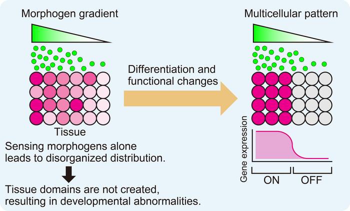
Advances in tissue culture techniques, particularly in the development of organoids and embryoids, have spurred interest in how tissues grow and organize in natural embryo development. While it is known that signaling molecules called morphogens drive tissue growth through diffusion, it has been a challenge to understand how these gradients lead to the formation of sharp boundaries between different tissue regions. Now, a research team led by Satoshi Toda (now at Osaka University), Kosuke Mizuno, and Tsuyoshi Hirashima have shed light on this process using a model called SYMPLE3D (Synthetic Morphogen system for Pattern Logic Exploration using 3D spheroids).
Previous studies focused separately on morphogens and cell adhesion proteins in tissue development. However, recent findings suggested that morphogens involved in neural tube development could regulate the expression of cadherins, a family of adhesion proteins, to create distinct tissue boundaries. Inspired by these insights, Toda and his team designed SYMPLE3D to investigate how morphogens and cadherins work together to form sharp boundaries in growing tissue.
The SYMPLE3D model utilizes two distinct cell types. One type, known as GFP secretors, releases GFP (Green Fluorescent Protein) and expresses P-cadherin, forming "GFP-secreting organizer spheroids." The second type, GFP receiver cells, are engineered to express a synthetic receptor called "synNotch" that recognizes GFP and activates a reporter gene, mCherry, in a process designed to mimic tissue patterning.
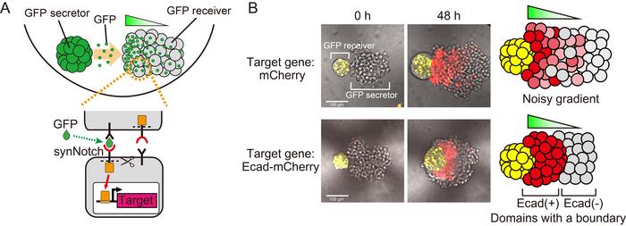
In the first stage of the experiment, the researchers co-cultured the GFP secretors and receiver cells. They observed a GFP gradient in the receiver cells, but the distribution was uneven, leading to the presence of cells expressing high levels of mCherry in inappropriate areas. To address this, the team engineered the GFP receiver cells to induce mCherry-fused E-cadherin, a cell adhesion molecule. This modification resulted in the formation of a uniformly activated tissue domain with a sharp boundary, rather than the previous gradient between the secretor and receiver cells.
The discovery was surprising: a single addition of E-cadherin drastically altered the patterning, creating a clear boundary that remained stable under different growth conditions. Toda and his team focused on understanding the mechanism behind this change. By tracking the tissue growth process in real-time, they found that initially scattered active cells expressing E-cadherin gradually aggregated, eventually absorbing the ectopically active cells into a single, sharply defined domain.
Further analysis revealed an intriguing detail—within the active domain, E-cadherin levels were uniformly high, despite GFP being distributed in a gradient. This observation highlighted a key role for E-cadherin in synthetic tissue domain formation. The researchers explored how cells behaved when expressing varying levels of E-cadherin in response to different GFP concentrations. Surprisingly, the cells' behavior was consistent, whether they expressed low or high levels of E-cadherin. Cells that expressed more than a certain threshold of E-cadherin were able to mix and form a unified population, regardless of their expression levels. This mixing allowed the cells to receive GFP evenly, which in turn led to uniformly high levels of E-cadherin expression across the synthetic tissue domain.
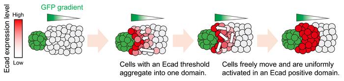
A simple mathematical model, developed by Hirashima, supported these experimental observations. Based on the principle of cell movement governed by differential adhesion energy, the model explained how cells with different E-cadherin levels could mix and form a coherent tissue structure with sharp boundaries.
"Our findings suggest the possibility of programming new tissue domains with sharp boundaries in organoids by combining synthetic morphogens with cell adhesion control," the team concluded. This research provides an exciting new tool for studying tissue patterning and could pave the way for advancements in organoid engineering and synthetic biology.
Read More


Newletter & More


SynBioBeta
Join the innovators shaping the future with SynBio + AI. From health to ag, materials & more—be part of the revolution.


