The Organoid Odyssey: A Leap Forward in Understanding Pediatric Brain Tumors
Scientists develop mini-brain organoids with unparalleled accuracy, potentially revolutionizing the study of cortical gliomas in children
Nov 29, 2023
AI Image Created Using DALL-E
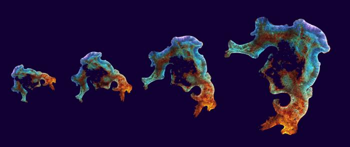
Let's embark on a cerebral journey where the marvels of science meet the intricacies of the human brain. Picture a group of visionary scientists, nestled in the heart of the Princess Máxima Center for Pediatric Oncology and the Hubrecht Institute, who have embarked on a groundbreaking endeavor. They've created something extraordinary: miniaturized versions of the human cortex, not just any replicas, but ones that echo the real deal with uncanny precision. These aren't your run-of-the-mill scientific models; they're organoids that mirror the human brain's architecture and cell identity, offering a new vista to explore pediatric brain tumors.
Imagine, if you will, a battlefield within the brain where childhood cortical gliomas, formidable foes, arise. The cortex, a bastion of complexity and the brain's most expanded structure is where these battles are waged. In this relentless fight, only about six in ten young warriors survive five years post-diagnosis with these malignant central nervous system tumors. But hope glimmers on the horizon as understanding the genesis and progression of these brain tumors could unveil new strategies for treatment.

Enter the world of organoids, where scientists cultivate 3D mini-organs and mini-tumors in the controlled environment of a laboratory. The Princess Máxima Center and the Hubrecht Institute have elevated this technique, creating a new cortex organoid that mirrors the human brain with unprecedented accuracy.
As reported recently in Nature Communications, Dr. Benedetta Artegiani and Delilah Hendriks stand at the forefront of this discovery. Their creation is a marvel, encapsulating the human brain's form, architectural organization, and cellular characteristics.
Let's delve into the genesis of our enigmatic brain, beginning with a singular structure, the neural tube composed of neuroepithelium. From this humble start, an intricate choreography unfolds, guiding cells to form the diverse regions of the brain.
Artegiani illuminates this complex dance, "The current brain organoid models generally have several developmental structures acting like an ‘independent’ small developing brain, within one organoid. Researchers have tried for a long time to mitigate the formation of these multiple structures. To diminish variability, increase reproducibility, better recreate the brain’s different cellular identities, and ultimately recapitulate brain development more closely. Now, in this research, the new organoid model is composed of this self-organizing, single developing neuroepithelium."

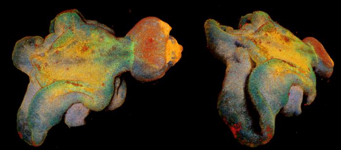
Anna Pagliaro, a PhD student, and the study's first author, reflects on their innovative approach, "We tried to mimic, in a dish, gradients of molecules that are present during brain development over time. This resulted in mini-brains that are very different in shape and structure than we used to work with. These organoids overall better resemble the early stages of brain development. It is quite impressive to see how much the shape of these organoids can influence all the different cells that form them, both in terms of their shape, architecture, but also identity."
These researchers have christened their creations Expanded Neuroepithelium Organoids (ENOs). A subtle yet impactful modification in their approach has been the gradual introduction of a molecule, TGF-b, mimicking the brain's development process.
Hendriks shares her insight, "Cells in the brain are instructed to acquire their identity through molecules that act slowly in time, the so-called temporal gradients. This is exactly what we tried to mimic. To our surprise, it was enough just to provide one of the molecules (TGF-b) slowly step-by-step to generate brain organoids. This tiny change had an enormous impact and allowed us to generate organoids with a shape and an identity more similar to the human brain."
The journey continues as these new organoid models offer a promising avenue for studying pediatric brain tumors, potentially tracing their origins to developmental anomalies. The researchers' focus on TFG-b signaling not only reflects its role in brain development but also its potential link to pediatric brain cancer.
Artegiani concludes with a vision, "We can use these novel organoids as a basis to model pediatric brain tumors and study more in depth the role of TFG-b signaling in this process. Our research marks an essential step to creating proper models to study pediatric brain cancer. If such a small change of a signaling molecule has such a great impact on brain organoid models, we can only start to imagine which effects small alterations during development can have for how pediatric brain tumors can develop."
This fusion of scientific rigor and creative exploration paves the way for a deeper understanding of one of life's greatest mysteries: the human brain.


